On this page
Why is follow-up necessary?
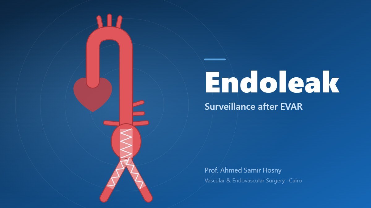
The old aneurysm sac remains around the graft. Ideally it slowly shrinks or stays stable. If blood leaks back into it, the sac may enlarge and the risk of rupture returns. This usually causes no symptoms, so only imaging can detect it.
Types of endoleak
- Type I: a leak at the upper or lower end of the graft because of incomplete sealing — usually needs prompt treatment.
- Type II: back-bleeding into the sac from small aortic branches (lumbar arteries or the inferior mesenteric artery) — the most common type; many seal spontaneously and are observed, and they are treated if the aneurysm grows.
- Type III: separation between graft components or a fabric tear — needs treatment.
- Type IV: minor seepage through the fabric pores right after the procedure; rare with modern grafts.
- Type V (endotension): sac enlargement without a visible leak on imaging.
The usual follow-up schedule
- A CT angiogram or duplex ultrasound about one month after the procedure.
- A scan at one year.
- Then usually yearly imaging, with duplex ultrasound and sometimes CT depending on the case and kidney function.
Your doctor may schedule closer scans if a leak or a change in sac size is found.
How are endoleaks treated?
Most endoleaks are treated with catheter techniques: adding an extension cuff or limb, extra fixation, or blocking the vessels feeding a type II leak with coils or medical glue. Open surgery is rarely needed.
When to seek urgent care
- Sudden, severe abdominal or back pain.
- Fainting or a drop in blood pressure.
- A cold, painful or discoloured leg, or new leg pain when walking.
- Unexplained fever after the procedure.
Frequently asked questions
Does an endoleak mean the operation failed?
Not necessarily. Many type II leaks are minor and resolve on their own, and the other types can usually be treated with a catheter procedure.
Can duplex ultrasound replace CT?
In many stable patients, yes — reducing radiation and contrast exposure. Your doctor chooses what suits your case.
Can I live a normal life?
Yes. Most people return to work and light exercise within a few weeks, provided they attend follow-up, control blood pressure and stop smoking.
A successful stent-graft needs successful follow-up — never postpone your surveillance scan.

Prof. Ahmed Samir Hosny
Professor of Vascular & Endovascular Surgery — Kasr Al Ainy, Cairo University
Professor of Vascular & Endovascular Surgery at Kasr Al Ainy, Cairo University. Fellowship in peripheral endovascular interventions (Kiel, Germany) and aortic surgery fellowship (Cologne, Germany); Member of the Royal College of Surgeons of Edinburgh.
Meet the doctor